Plasmodial Slime Mold Consuming A Trametes Fungus By Michael Harz

plasmodial slime mold consuming a Trametes fungus by Michael Harz
More Posts from Mikrobiotch and Others
Microbiology!

[ID: a banner made of emojis of microscopes, bubbling flasks, and DNA, with different bacteria emojis from a combo emoji scattered between them. /End ID]


Stemonitis sp.
photo source-The MacroClub Project (Myxomycetes)
Slime Mold

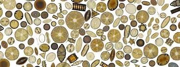
Coelastrum, a microalgae.


These pictures show PAS (purple/pink) and GMS (brown/blue) staining of a lymph node biopsy from a canine patient with lymphadenopathy and weight loss. Histology was suggestive of likely a fungal organism (bright pink in the PAS stain and dark brown/black in the GMS stain), however a mixed infection with an algal species could not be definitively ruled out without microbiology.
Our microbiologist cultured an Aspergillus species from this dog, and is in the process of ruling out any other possibilities.
What's especially cool is in one of the PAS pictures, you can see an organism trapped within an actively dividing macrophage!


Craterellus cornucopioides (trumpet of the dead) and Hygrocybe conica (witch's hat), competing for Most Goth Common Name

DNA from a strawberry!!! This was super cool (the little white strands in the clear is actual dna from a strawberry!) strawberries are octoploids which means they have 8 copies of each chromosome! It makes it easier to see and extract it’s DNA. That’s wild!


Scientists from BGI-Research developed a new version of the Cultivated Genome Reference (CGR), a repository of high-quality draft genomes of the human gut microbiome. The current version of CGR, which is CGR2, has been further expanded to incorporate numerous high-quality draft genomes generated from cultivated bacteria. CGR2 classifies previously unidentified species and uncovers the functional and genomic diversity of bacterial strains. An in-depth analysis of carbohydrate-active enzymes (CAzymes) reveals the phyla with the largest and most diverse repertoires of these enzymes. CGR2 also enabled the identification of genes involved in the synthesis of secondary metabolites in the gut microbiome. The unraveling of the gut microbiome genomic landscape will enable the development of therapeutics and provide a deep insight into the evolution of the human gut microbiome.
Continue Reading
Watch what happens to Germs when you wash your hands with Soap at microscopic level. 🔬 The Soap molecules surround germ cells and disrupt their cell walls, causing them to burst.
Germ cells are surrounded by a cell wall that protects them from the environment. This cell wall is made up of a layer of peptidoglycan, which is a polymer of amino acids and sugars. Soap molecules are made up of two parts: a hydrophobic (water-fearing) tail and a hydrophilic (water-loving) head. When soap is added to water, the hydrophobic tails group together and the hydrophilic heads face outward, forming micelles. These micelles can surround germ cells and the hydrophobic tails can then disrupt the cell walls, causing the cells to burst.
The hydrophobic tails of the soap molecules can disrupt the cell wall in two ways. First, they can bind to the peptidoglycan molecules and weaken the bonds between them. Second, they can create holes in the cell wall. Once the cell wall is disrupted, the germ cells lose their internal contents and die.
It is important to note that soap only works to kill germ cells that are surrounded by a cell wall. Germ cells that do not have a cell wall, such as viruses, are not affected by soap.
The size of the soap micelles is important. Micelles that are too small will not be able to surround the germ cells. Micelles that are too large will not be able to penetrate the cell walls.
The concentration of soap is also important. A higher concentration of soap will be more effective at killing germ cells.
The temperature of the water can also affect the effectiveness of soap. Soap is more effective at killing germ cells in warm water than in cold water.
I hope this post has helped you understand the importance of handwashing and why doctors always ask you to do it regularly. Washing your hands with soap and water for at least 20 seconds is one of the best ways to prevent the spread of germs and stay healthy. So please, wash your hands often and help keep yourself and others safe!
Thank you for reading this post. I hope you found it informative and helpful. Please share it with your friends and family so they can learn about the importance of handwashing too. 😊🙏
FOTD #071 : red coral fungus! (ramaria araiospora)
red coral is a coral mushroom in the family gomphaceae. :-) it is found in the himalaya & north america. it grows either in clusters or singularly, & prefers western hemlock & tanoak. it likely forms a mycorrhizal association !!
the big question : can i bite it?? it is edible & sold as food in mexico :-) though, overconsumption can cause stomach upset.


r. ariospora description :
"the fruit bodies of ramaria araiospora typically measure 5–14 cm (2–5+1⁄2 in) tall by 2–10 cm (3⁄4–3+7⁄8 in) wide. there is a single, somewhat bulbous stipe measuring 2–3 cm (3⁄4–1+1⁄8 in) long by 1.5–2 cm (5⁄8–3⁄4 in) thick, which is branched up to six times. the branches are slender, usually about 1–5 mm (1⁄16–3⁄16 in) in diameter, while branches near the base are thicker, up to 4 cm (1+5⁄8 in) thick. the terminal branches are forked or finely divided into sharp tips. the trama is fleshy to fibrous in young specimens, but becomes brittle when dried. the branches are red initially, fading to a lighter red in maturity, while the base, including the stipe, is white to yellowish-white. branch tips are yellow."
[images : source & source] [fungus description : source]
"i love this fungus so much<3 she's SO pretty. i only learnt about it recently."
-
 annoyinglyeclecticruins reblogged this · 3 months ago
 -
annoyinglyeclecticruins liked this · 3 months ago
-
 dinosaurwithablog liked this · 6 months ago
 -
 mycellpics liked this · 1 year ago
 -
 tsubami612 reblogged this · 1 year ago
 -
tsubami612 liked this · 1 year ago
-
 corrodedcarcass liked this · 1 year ago
 -
 therussianbudgie liked this · 1 year ago
 -
 jamie0721 reblogged this · 1 year ago
 -
 allhailthedumpsterfire reblogged this · 1 year ago
 -
allhailthedumpsterfire liked this · 1 year ago
-
 uglyluckylucky reblogged this · 1 year ago
 -
 bprodukt liked this · 1 year ago
 -
 unofficial-sean reblogged this · 1 year ago
 -
unofficial-sean liked this · 1 year ago
-
 whereismarilyn reblogged this · 1 year ago
 -
 dave-of-the-rave liked this · 1 year ago
 -
 sludgedevine liked this · 1 year ago
 -
 kyoshx reblogged this · 1 year ago
 -
 gringatrash reblogged this · 1 year ago
 -
 gravediggers reblogged this · 1 year ago
 -
 verminhost reblogged this · 1 year ago
 -
 namorailecec liked this · 1 year ago
 -
 mariposasmonarch liked this · 1 year ago
 -
 rostii liked this · 1 year ago
 -
 trans-elrond liked this · 1 year ago
 -
 strongcat liked this · 1 year ago
 -
 pvrrhadve reblogged this · 1 year ago
 -
 the-name-recs-are-crazy reblogged this · 1 year ago
 -
rostii reblogged this · 1 year ago
-
 evilgoatsimulator liked this · 1 year ago
 -
 homeplanets reblogged this · 1 year ago
 -
 dhdiqnd1 liked this · 2 years ago
 -
 gblahlife liked this · 2 years ago
 -
 dontknowanymorethanyoudo liked this · 2 years ago
 -
 naturalblondekiller reblogged this · 2 years ago
 -
naturalblondekiller liked this · 2 years ago
-
 honeybadgersfandom liked this · 2 years ago
 -
 campfema liked this · 2 years ago
 -
 spacefinch liked this · 2 years ago
 -
 tree-whisper liked this · 2 years ago
 -
 sztefa001 reblogged this · 2 years ago
 -
 mbhfisme reblogged this · 2 years ago
 -
 1568644byeee reblogged this · 2 years ago
